¡Bravo! 23+ Hechos ocultos sobre Anatomy Chest Muscles Diagram? Start studying chest muscles anatomy.
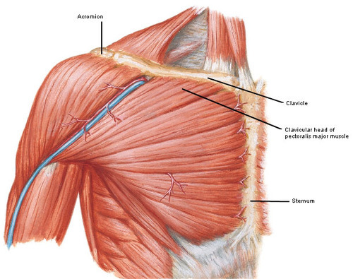
Anatomy Chest Muscles Diagram | Try these free muscle labeling diagrams. For example, some muscles located in the chest also help move the shoulders. Pectoral muscles are the muscles that connect the front of the human chest with the bones of the upper arm and shoulder. Muscles make up a large part of the anatomy (structure) of the back. The pectoralis major is the superior most and largest muscle of the anterior chest wall.
And receive our free ebook: Start studying chest muscles anatomy. It's innervated by both medial and lateral pectoral nerves. Try these free muscle labeling diagrams. Deep muscles of the chest, .

Each breast consists of tissue overlying the chest wall muscles (the pectoral muscles). The pectoralis major is a prominent chest muscle that acts mainly on the shoulder joint. Pectoral muscles are the muscles that connect the front of the human chest with the bones of the upper arm and shoulder. The medical name for breast is mammary gland. Try these free muscle labeling diagrams. Deep muscles of the chest, . It's innervated by both medial and lateral pectoral nerves. Learn the anatomy of the pecs now at kenhub! Start studying chest muscles anatomy. There are two such muscles on . Guide to mastering the study of anatomy. Learn vocabulary, terms, and more with flashcards, games, and other study tools. And receive our free ebook:
Try these free muscle labeling diagrams. There are two such muscles on . And receive our free ebook: Learn vocabulary, terms, and more with flashcards, games, and other study tools. Each breast consists of tissue overlying the chest wall muscles (the pectoral muscles).

Each breast consists of tissue overlying the chest wall muscles (the pectoral muscles). Learn the anatomy of the pecs now at kenhub! The dominant muscle in the upper chest is the pectoralis major. Deep muscles of the chest, . Try these free muscle labeling diagrams. Learn vocabulary, terms, and more with flashcards, games, and other study tools. Pectoral muscles are the muscles that connect the front of the human chest with the bones of the upper arm and shoulder. Pectoralis muscle, any of the muscles that connect the front walls of the chest with the bones of the upper arm and shoulder. And receive our free ebook: Muscles make up a large part of the anatomy (structure) of the back. The pectoralis major is a prominent chest muscle that acts mainly on the shoulder joint. It's innervated by both medial and lateral pectoral nerves. The pectoralis major is the superior most and largest muscle of the anterior chest wall.
Pectoral muscles are the muscles that connect the front of the human chest with the bones of the upper arm and shoulder. Deep muscles of the chest, . The dominant muscle in the upper chest is the pectoralis major. Muscles make up a large part of the anatomy (structure) of the back. Pectoralis muscle, any of the muscles that connect the front walls of the chest with the bones of the upper arm and shoulder.
The muscles of the chest and upper back occupy the thoracic region. For example, some muscles located in the chest also help move the shoulders. And receive our free ebook: It's innervated by both medial and lateral pectoral nerves. The pectoralis major is a prominent chest muscle that acts mainly on the shoulder joint. Try these free muscle labeling diagrams. Pectoralis muscle, any of the muscles that connect the front walls of the chest with the bones of the upper arm and shoulder. The medical name for breast is mammary gland. Muscles make up a large part of the anatomy (structure) of the back. Learn vocabulary, terms, and more with flashcards, games, and other study tools. There are two such muscles on . Pectoral muscles are the muscles that connect the front of the human chest with the bones of the upper arm and shoulder. Start studying chest muscles anatomy.
Anatomy Chest Muscles Diagram! Learn the anatomy of the pecs now at kenhub!
0 Response to "¡Bravo! 23+ Hechos ocultos sobre Anatomy Chest Muscles Diagram? Start studying chest muscles anatomy."
Post a Comment